Posts

Showing posts from January, 2013

Single-File Technique Sample 2

Image
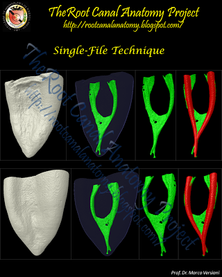
Click here to download an editable Power Point file with all pictures and movies of the single-file technique series Mesial root of mandibular molar  -   The 3D model in  GREEN  indicates the  original root canal anatomy . The 3D model in  RED  indicates the root canal modified  after preparation  with a single-file system superimposed to the  original canal .

Single-File Technique Sample 1

Image
Single-File Technique Series The introduction of nickel-titanium (NiTi) rotary file systems has resulted in a markedly progress on the mechanical preparation of the root canal space. NiTi instruments offer many advantages over conventional stainless steel files such as flexibility and cutting efficiency. Furthermore, NiTi instruments maintain the original canal shape during preparation and have reduced tendency to transport the apical foramen. However, the current technology for mechanical preparation - rotary NiTi files - have failed in debriding oval-shaped canals, leaving untouched fins or recesses on the buccal and/or lingual extensions. This untouched recesses may harbor unaffected residual bacterial biofilms and serve as a potential cause of persistent infection and poor treatment outcome. Besides, as these techniques also require the use of numerous instruments to enlarge the canal to an adequate size and taper, they are relatively time and cost consuming. The newly developed rec...

Single-File Technique Sample 6

Image
Click here to download an editable Power Point file with all pictures and movies  of the single-file technique series Mesial root of mandibular molar  -   The 3D model in  GREEN  indicates the  original root canal anatomy . The 3D model in  RED  indicates the root canal modified  after preparation  with a single-file system superimposed to the  original canal .

Single-File Technique Sample 5

Image
Click here to download an editable Power Point file with all pictures and movies  of the single-file technique series Mesial root of mandibular molar  -   The 3D model in  GREEN  indicates the  original root canal anatomy . The 3D model in  RED  indicates the root canal modified  after preparation  with a single-file system superimposed to the  original canal .

Single-File Technique Sample 4

Image
Click here to download an editable Power Point file with all pictures and movies of the single-file technique series Mesial root of mandibular molar  -   The 3D model in  GREEN  indicates the  original root canal anatomy . The 3D model in  RED  indicates the root canal modified  after preparation  with a single-file system superimposed to the  original canal .

Single-File Technique Sample 3

Image
Click here to download an editable Power Point file with all pictures and movies of the single-file technique series Mesial root of mandibular molar  -   The 3D model in  GREEN  indicates the  original root canal anatomy . The 3D model in  RED  indicates the root canal modified  after preparation  with a single-file system superimposed to the  original canal .

Links for downloading

Image
ANNOUNCEMENT Dear readers , This weekend I realized that there were several missed links for downloading the material from The Root Canal Anatomy Project blog. I am sorry if it caused you some disappointment. I would like to announce that all missed links were corrected and the material is able to be downloaded again. Please, if you detect some problems in this blog do not hesitate in contacting me by email: marcoversiani@yahoo.com Thanks a lot! Enjoy, Prof. Marco Versiani, DDS, MS, PhD

Root Canal Preparation Systems in Endodontics

To download a PDF file of this presentation CLICK HERE Mechanical Preparation Systems in Endodontics from Prof Dr. Marco Versiani

Self-Adjusting File: Article

Image
 For more information, click HERE . Abstract  Introduction Cleaning and shaping of root canals are essential steps for the success of endodontic therapy. The purpose of this study was to evaluate the tissue debridement efficacy of the self-adjusting file (SAF) protocol in the apical third of oval-shaped canals of mandibular incisors in comparison with a nickel-titanium rotary system preparation. Methods Twenty-six single-rooted human mandibular incisor teeth were selected and assigned to a control (n = 4) and 2 experimental groups (n = 11) according to 1 of 2 instrumentation techniques, SAF and nickel-titanium rotary systems. After root canal preparation, the apical thirds of the specimens were submitted to histologic processing and analyzed by optical microscopy regarding the percentage of debris and uninstrumented root canal walls. The data were statistically compared by using unpaired t test with Welch's correction, and the level of significance was set at 5%. Results The p...

Mandibular Canine

Image
The article entitled " Microcomputed tomography study of the root canal morphology of single-rooted mandibular canine " was published on the  International Endodontic Journal . By now, I am going to provide you with some pictures and a movie of this group of teeth.  P.S. The original article contains different pictures from the below. Clique HERE to download an editable Power Point file with all pictures and the movie below

Enamel Pearls in Permanent Dentition

Image
The article entitled " Enamel pearls in permanent dentition: case report and micro-CT evaluation " was published on the  Dentomaxillofacial Radiology  journal. By now, I am going to provide you with some important information about this morphological variation of teeth.  P.S. The original article contains different pictures from the below. The following text was extracted from: Moskow BS, Canut PM. Studies on root enamel (2). Enamel pearls. A review of their morphology, localization, nomenclature, occurrence, classification, histogenesis and incidence. J Clin Periodontol 1990; 17: 275–281. Surface Description When examined grossly or macroscopically, enamel pearls most commonly appear spheroid, however other shapes such as conical, ovoid, tear-drop, cylindrical and irregular have been observed (Negra & Olliveira 1974). They vary in size from a pine-head or smaller to that of a large cusp of a tooth. In one study of over 7000 teeth, the mean diameter of these enamel st...

Chile 2013

Image

A Travel Through a Maxillary Central Incisor

Image
Click here to download this video