Posts

Showing posts with the label Canal Preparation

XP-endo Shaper and XP-endo Finisher

Image
Preparation of curved canals of a mandibular first molar with XP-endo Shaper and XP-endo Finisher Prof. Marco Versiani, Prof. Kleber Carvalho & Prof. Sousa Neto

Hard Tissue Debris Removal: XP Endo Finisher

Image

Rotary Preparation: Mandibular Molar

Image
Click here to download an editable Power Point file with all pictures and video

Root Canal Anatomy X Rotary Preparation

Image
The influence of the anatomical configuration of the root canal on the root canal preparation Type IV (Vertucci's Configuration System) Type V (Vertucci's Configuration System) Watch this video in HD!

Root Canal Preparation: Mandibular Molar

Image
Root Canal Preparation of a Mandibular Molar  Using a Multiple-File Rotary NiTi System Thanks to Dr. Ali Keles from Turkey for the scanning and canal preparation procedures In green : root canal before preparation In red : root canal after preparation superimposed to the original canal Click here to download an editable Power Point file with all pictures and movie

Single-File Technique Sample 2

Image
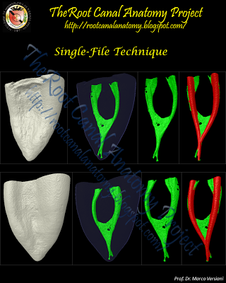
Click here to download an editable Power Point file with all pictures and movies of the single-file technique series Mesial root of mandibular molar  -   The 3D model in  GREEN  indicates the  original root canal anatomy . The 3D model in  RED  indicates the root canal modified  after preparation  with a single-file system superimposed to the  original canal .

Single-File Technique Sample 1

Image
Single-File Technique Series The introduction of nickel-titanium (NiTi) rotary file systems has resulted in a markedly progress on the mechanical preparation of the root canal space. NiTi instruments offer many advantages over conventional stainless steel files such as flexibility and cutting efficiency. Furthermore, NiTi instruments maintain the original canal shape during preparation and have reduced tendency to transport the apical foramen. However, the current technology for mechanical preparation - rotary NiTi files - have failed in debriding oval-shaped canals, leaving untouched fins or recesses on the buccal and/or lingual extensions. This untouched recesses may harbor unaffected residual bacterial biofilms and serve as a potential cause of persistent infection and poor treatment outcome. Besides, as these techniques also require the use of numerous instruments to enlarge the canal to an adequate size and taper, they are relatively time and cost consuming. The newly developed rec...

Single-File Technique Sample 6

Image
Click here to download an editable Power Point file with all pictures and movies  of the single-file technique series Mesial root of mandibular molar  -   The 3D model in  GREEN  indicates the  original root canal anatomy . The 3D model in  RED  indicates the root canal modified  after preparation  with a single-file system superimposed to the  original canal .

Single-File Technique Sample 5

Image
Click here to download an editable Power Point file with all pictures and movies  of the single-file technique series Mesial root of mandibular molar  -   The 3D model in  GREEN  indicates the  original root canal anatomy . The 3D model in  RED  indicates the root canal modified  after preparation  with a single-file system superimposed to the  original canal .

Single-File Technique Sample 4

Image
Click here to download an editable Power Point file with all pictures and movies of the single-file technique series Mesial root of mandibular molar  -   The 3D model in  GREEN  indicates the  original root canal anatomy . The 3D model in  RED  indicates the root canal modified  after preparation  with a single-file system superimposed to the  original canal .

Single-File Technique Sample 3

Image
Click here to download an editable Power Point file with all pictures and movies of the single-file technique series Mesial root of mandibular molar  -   The 3D model in  GREEN  indicates the  original root canal anatomy . The 3D model in  RED  indicates the root canal modified  after preparation  with a single-file system superimposed to the  original canal .

Root Canal Preparation: Gates-Glidden + K-Files

Image
Root Canal Preparation Protocol (Crown-Down Technique): Mesiobuccal and Mesiolingual Canals  - Cervical Third (Gates-Glidden burs #3 - #1) - Middle and Apical Thirds (Stainless Steel K-Files - Apical Preparation: K-File: #30) Distal Canal - Cervical Third (Gates-Glidden burs #2 - #4) - Middle and Apical Thirds (Stainless Steel K-Files - Apical Preparation: K-File: #40) Click on the images to enlarge 3D Models of Root Canal Before Preparation ( Green ) 3D Models of Root Canal After Preparation ( Red ) 3D Models of Root Canal Before and After Preparation Registration Process Five Transaxial Views Of The Root Superimposition of the Canal Before and After Preparation Superimposition of the 3D Models of the Root Canal Before ( Green ) and After ( Red ) Canal Preparation Cervical Preparation With Gates-Glidden Burs Before (Black) and After ( Red ) Canal Preparation Axial Cross-Sections of a Mandibular Molar Root Canal Preparation (Crown-Down Technique) [Gates-Glidden Burs + Stainless...

Single-File Preparation

Image
"Shape most root canals using only one file and decrease total shaping time..." I am sorry, but in cases like this one, the total shaping time should be increased to allow an adequate disinfection of the isthmuses that were not even touched by the instrument. Otherwise, most of the canal surface area will be kept without any treatment. With these images, it is possible to understanding the real influence of the root canal anatomy on the endodontic procedures. The root canal space is an arborizational, anastomotic, labyrinthine complexity, morphologically comparable to the pathways of a maze. While primary canals exist, the tributaries, accessory branches and lumina of the dentinal tubuli harbor extensive tissue and microflora. The existence of these vast, capacious passages has been demonstrated throughout the past century beginning with the work of Hess and continues to this day with the use of micro computed tomography. Presence of multiple foramina, additional canals...